Root Canal Treatment
A root canal might sound scary but it’s a fairly standard dental procedure performed on millions of Americans every year. Our team of dental specialists will explain the importance of root canal treatment and how they will help you in maintaining a happy, healthy smile.
When is a Root Canal Procedure Needed?
A root canal can be indicated when suffering from a cracked tooth, when tooth decay has spread to the nerve, trauma, or when causing extreme sensitivity due to a deep filling.
Other symptoms signaling a need for a root canal:
- Pain while eating or spontaneous constant pain
- Chipped/ cracked tooth
- Swelling in the gums
- Prolonged sensitivity to hot and cold fluids
- Injury to the tooth
What is the Treatment Procedure for a Root Canal?
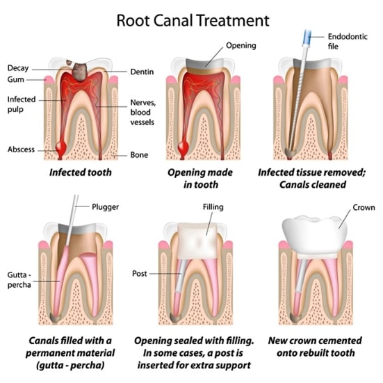
The process comprises of following steps-
- Diagnosis of the infected tooth
- Pulp removal
- Replacing the pulp tissue with a rubber compound called gutta-percha
- Crown placement

Root Canal Recovery
- Post root canal procedure, the mouth will remain numb for 2-3 hours maximum.
- You can resume office, college or other activities post procedure.
- Wait for the numbness to wear off before drinking or eating.
- Soreness can occur in the first 1-3 days after an endodontic treatment. Manage with Tylenol or Ibuprofen.
- Avoid chewing food from the treated site till the root canal procedure is completed.
- The brushing and flossing routine remains the same as before
- If experiencing severe pain that can’t be managed with OTC pain meds, please call your dentist for a follow up evaluation.

Benefits of a Root Canal
- Relieving tooth pain.
- Retaining natural teeth.
- Preventing Jawbone degeneration.
- Prevention of possible future dental implants procedure.
- Pain-free consumption of hot or cold fluids and solid food.
- Value for money.
- Visually appealing.
Root Canal Cost
Treatment cost depends on multiple factors like insurance, the specific tooth requiring treatment, the clinic’s location, and the dentist’s expertise, but the average cost of treatment is typically around $1,020.
Specialized Equipment
First Point Dental is equipped with state-of-the art technology to deliver the highest quality of care to their patients. Take a look at the different equipment available for root canal treatment:
Digital Radiography (X-Rays)
- Our dental clinics are equipped with modern digital x-ray equipment. Your digital x-rays will be examined on a high-resolution digital screen. Plus, the tooth can be x-rayed with less radiation. In our clinic, each digital x-ray is just 1/10th of the normal radiation exposure of the traditional dental x-ray.
- Today, Digital x-ray technology provides better resolution than traditional methods. This helps the dentists in getting better diagnostic accuracy.
- By enhancing the x-ray image, the dentist can explain the problem clearly and explain the treatment needed to save the teeth.
- Images of infected teeth from your x-rays are stored confidentially on our secure database. In future follow-ups and oral treatment, we will have a picture of the progression or decline of the infection and decay.
Surgical Microscopes and Endoscopes
Our dental offices are equipped with advanced surgical microscopes and digital endoscope equipment. This is advantageous in diagnosing and treating difficult dental cases.
What it does:
- Locating calcified or blocked canals,
- Identifying tooth’s micro-fractures
- Retrieving broken fragments of the instruments from the root canal
- Identifying perforation of the tooth’s internal surface
Cone Beam Computerized Tomography (CBCT)
- At First Point Dental, we believe in the idea of offering the best-specialized service. A 3-D Cone Beam CT (CBCT) allows us to do just that!
- With this tool, our specialists can produce the highest resolution imaging to correctly diagnose and provide the corrective response to treatment.
- The 3-D cone beam images are more accurate and complete with negligible radiation. 3-D cone beam images can be copied and stored for future reference.
- The doctors have access to powerful, focused, 3D images, and a high-resolution x-ray of the oral cavity.
- Complete visualization of lesions and anatomy
- Access unseen anatomical detail to correctly diagnose the problem
- Clear visualization of tooth canals as well as identifying the lesions source
